J Med Discov (2024); 9(1): jmd24026; DOI:10.24262/jmd.9.1.24026;
Received January 20th, 2024, Revised March 08th, 2024, Accepted March 15th, 2024, Published March 25th, 2024.
Observation on the Efficacy of Layered Fat Grafting via Intraoral Channel in Nasolabial Fold Aging
Lina Yang, Enfeng Wang, Zhuomao Xu, Jingdan Fu,Yiqun Sun*
1Department of Plastic and Burn, Hainan Western Central Hospital, Danzhou 571700, China
* Correspondence:Yiqun Sun, Department of Plastic and Burn, Hainan Western Central Hospital, Danzhou 571700, China.Email: sun-yiqun@163.com
Abstract
Objective This study aimed to explore the method and efficacy of using small granular fat and SVF-gel through intraoral channel for the treatment of nasolabial fold aging in women. Methods A total of 20 patients (aged 25-68 years, mean age 46 years) with nasolabial fold aging were selected from the Department of Plastic and Burn Surgery at Hainan Western Central Hospital from October 2021 to April 2023. Preoperative assessment of the severity of nasolabial fold wrinkles was performed using the Wrinkle Severity Rating Scale (WSRS). The surgical technique involved layered transplantation of small granular fat grafts on the periosteal layer and SVF-gel on the subcutaneous layer through an intraoral channel. Results None of the 20 patients experienced complications such as local induration, infection, pain, swelling, or bruising after the surgery. All patients achieved a smooth or slightly elevated appearance of the nasolabial fold immediately after the surgery, and the immediate effect is good, with satisfactory outcomes during follow-up visits ranging from 1 to 12 months. Conclusion Layered fat grafting via intraoral channel for the treatment of nasolabial fold aging in women offers advantages such as minimal intraoperative bleeding, low incidence of postoperative complications, and no visible scarring on the facial skin. The high level of patient satisfaction indicates the potential for widespread adoption of this technique.
Keywords:Nasolabial fold aging; Granular fat; SVF-gel; Intraoral channel; Layered transplantation.
The anatomical layers of the nasolabial fold region are divided into skin layer, fat layer, superficial musculoaponeurotic system (SMAS), fibrous junction layer, and muscle layer. Nasal base refers to the basal part of the nose connected to the upper lip, that is, the bony site centered on the anterior nasal crest and extending to both sides. The nasal base depression makes the nasolabial fold in the middle of the face obvious [1]. Autologous fat grafting to fill nasolabial fold is a commonly used clinical treatment in recent years [2]. Previous basic experiments have found that [3] stromal vascular fraction gel (SVF-gel) can significantly improve the survival rate of fat. SVF-gel removes large adipocytes in granular fat by physical methods, and enriched more adipose derived stem cells (ADSCs) and extracellular matrix (ECM) components [4]. From October 2021 to April 2023, 20 patients with nasolabial fold aging were admitted to the Department of Plastic and Burn Surgery, Hainan West Central Hospital. The fat layer filling through the intraoral channel was used to treat the nasolabial fold aging, and the effect was good. It is reported below.
1. Materials and Methods
1.1 General data
There were 20 patients in this group. All patients were female. The mean age was (46.2±8.9) years (range, 25-68 years). Before operation, the wrinkle severity rating scale (WSRS) was used to grade the severity of nasolabial fold wrinkles: 8 cases were 2 points and 12 cases were 3 points.
Inclusion criteria: ⑴ Nasolabial fold depression was diagnosed according to facial symptoms and examination. ⑵ Female, aged 18-70 years old. ⑶ Indications for soft tissue tamponade were found. ⑷ The WSRS score was 2-4 points. ⑸ High compliance and complete clinical data. ⑹ Bilateral nasolabial fold symmetry, equal filling volume. He was willing to be followed up after surgery.
Exclusion criteria: ⑴ Did not meet the diagnostic criteria of nasolabial fold, pathological diseases caused by nasolabial fold deepening. ⑵ The severity difference between the two sides of the nasolabial fold is too large. ⑶ Patients who had received nasolabial fold rejuvenation treatment. ⑷ serious diseases of organs and systems are not suitable for surgery and chronic diseases are not controlled (such as high blood pressure and high blood glucose). ⑸ Patients undergoing anticoagulant therapy, taking anticoagulant and blood-activating drugs, and anyone with bleeding tendency. ⑹ Facial skin with serious damage, infection, malignant lesions. He has severe mental illness and no behavioral capacity (such as severe anxiety and depression). ⑻ Pregnancy, lactation and the recent period of time to prepare a pregnancy plan. ⑼ Patients with scar constitution. ⑽ Preoperative routine examination (blood routine, six coagulation tests, pre-transfusion screening, liver and kidney function, electrocardiogram, chest X-ray) have serious abnormalities.
1.2 Surgical methods
⑴ Preoperative preparation. Preoperative medical history and physical examination were performed to screen for severe diseases, associated infections, bleeding tendencies, and allergies. Drugs containing aspirin and non-steroidal anti-inflammatory drugs were discontinued 2 weeks before surgery, and certain traditional Chinese medicine combinations that promote anticoagulation were discontinued. Prophylactic antibiotics were used before operation. ⑵ Donor site preparation and fat extraction. The patient was placed in the supine position, and after the operation area was disinfected and covered with towels, the skin near the inner thigh of the groin was used as the needle entry mark point, and the suction range was marked. After local anesthesia with 2% lidocaine 0.1 ml at the needle marking point, swelling anesthesia was performed with 0.1% lidocaine 100 ml in the marked suction area, and subcutaneous swelling solution was injected until the local skin became hard, white, and slightly elevated. The skin was punctured with a sharp knife, and liposuction was performed manually with a 2.5 mm liposuction needle connected to a 10 ml syringe under negative pressure. About 40 ml of fat was slowly aspirated. ⑶ Preparation of granular fat and SVF-gel. The original method was improved. The fibrous connective tissue and other impurities were removed with forceps, and the upper oil and the lower bloody liquid were removed by centrifugation at 200×g for 1 min to obtain granular fat. The obtained granular fat 10 ml was reserved, and the remaining 20 ml fat was used to prepare SVF-gel. The samples were centrifuged at 1500×g for 3 min. The midlayer adipose tissue was transferred to a sterile 10-ml syringe. The cells were connected with another 10 ml sterile syringe with a cutting connection head, and the piston of the syringe was rapidly pushed back and forward 20 times, and the negative pressure generated by reverse traction was used to rapidly rupture the cells. This was reloaded into a centrifuge and centrifuged at 2000×g for 2 min. It can be seen that the middle layer tissue is gelatinous, namely SVF-gel[13]. (4) Nasolabial fold filling. After routine facial disinfection, the upper lip was opened and the oral cavity was disinfected again, and the mucosa was punctured at the vestibular sulcus corresponding to the first upper left premolar and the first upper right premolar. An 18 G fat injection needle was inserted into the nasal base through the gingival sulcus, and the granular fat was injected into the upper periosteum of the nasolabial fold according to the volume of the nasolabial fold depression. After filling and smooth, the injection volume was added 20%. SVF-gel was injected into the deep dermis and subendothelial layer of the nasolabial fold with a 1 ml syringe connected to a 27 G fat injection needle. After the operation, the wound in the mouth was rinsed with low concentration of antibiotics and saline, and then the wound was closed by applying Fuaile medical glue (Beijing Fuaile Technology Development Co., LTD.). The volume of granule fat injection was about 3.0 ml/ side, and the volume of SVF-gel injection was about 0.5 ml/ side.
1.3 Criteria for efficacy evaluation
WSRS was used to score the severity of the nasolabial fold before and after surgery, which was divided into five grades from none to extremely severe, as shown in Table 1.
Table 1 Nasolabial Fold Severity Rating Scale (WSRS)
| Score | Performance |
| None (1 point) | No folds were seen, only continuous skin lines. |
| Mild (2 points) | Showed shallow folds and skin depression, and fine wrinkles. |
| Moderate(3points) | Wrinkles were deep and clear, but disappeared when stretched. |
| Severe(4 points) | Wrinkles were deep and long, obvious, and less than 2 mm visible wrinkles during extension. |
| Extreme(5 points) | Folds, very long, stretching from time to tome 2 ~ 4 mm clear Zha V visible wrinkles. |
1.4 Statistical treatment
SPSS 19.0 software was used to analyze the data. The measurement data were expressed as , and the comparison before and after operation was analyzed by two-sided t test, and P < 0.05 was considered statistically significant.
2 Results
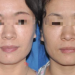
Twenty patients had smooth appearance of nasolabial fold immediately after operation. There were no complications such as local induration, infection, pain, swelling and cyanosis. The patients were followed up for 6 to 12 months, and the nasolabial fold was smooth at the last follow-up. See Figures 1,2. WSRS score at the last follow-up: 8 patients with preoperative WSRS score of 2 points, postoperative WSRS score of 1 point; Of the 12 patients with preoperative score of 3, 10 patients had postoperative score of 2, and 2 patients had postoperative score of 1. The difference of WSRS scores before and after surgery was statistically significant (P < 0.05), as shown in Table 2.
Table 2 Comparison of WSRS scores before and after surgery (score, )
| Time(n=20) | WSRS Score |
| Before surgery | 2.6±0.3 |
| After surgery | 1.5±0.4 |
| T value | 15.561 |
| P value | <0.05 |
Figure 1 A 42-year-old woman with intraoral granular fat upper periosteal filling +SVF-gel deep dermal, subendothelial filling. Filling volume: 3.0 ml/ side of granular fat; SVF-gel 0.5 ml/ side a. Before surgery b. 12 months after surgery
Figure 1 A 42-year-old woman with intraoral granular fat upper periosteal filling +SVF-gel deep dermal, subendothelial filling. Filling volume: 3.0 ml/ side of granular fat; SVF-gel 0.5 ml/ side a. Before surgery b. 12 months after surgery
3 Discussion
The morphological changes of the nasolabial fold can be caused by physiological or pathological changes at any level. Pronounced nasolabial fold deepening is caused by a combination of sagging and thinning of the facial skin and selective fat deposition outside the wrinkles. Skin aging is characterized by the gradual decrease of hyaluronic acid, collagen, elastin and other components in the dermis, and the skin gradually shows dryness and roughness, followed by the formation of deepening wrinkles. The shear force generated by the relative movement of skin, subcutaneous tissue and superficial musculoaponeurial system around the nasolabial fold caused by long-term facial expression muscle activity can cause the deepening of the nasolabial fold wrinkles [5]. The malar fat pad was limited to sag below the nasolabial fold due to the attachment of the expression muscle group at the nasolabial fold skin. In the process of aging, nasolabial fold wrinkles become more obvious due to the gradual atrophy of midfacial deep fat, displacement of superficial fat, and sagging of malar fat pad [6-7]. In addition, maxillary atrophy or congenital dysplasia can retract the upper lip tissue and aggravate nasolabial fold wrinkles [8-9].
At present, the methods to improve the nasolabial fold mainly include surgical treatment and non-surgical treatment. The surgical treatment is mainly midface lifting, and the non-surgical treatment is mainly injection filling. The commonly used filling injection materials include hyaluronic acid and autologous fat. Due to the large trauma, obvious scar, and long recovery time of surgical treatment, patients are often willing to accept non-surgical treatment, and expect to achieve good improvement results by convenient, minimally invasive, and non-trace methods. Hyaluronic acid has a short action time, requires repeated injections, and has safety problems. Autologous fat is a permanent filling material derived from itself. Due to its rich source, simple sampling, no allergic rejection, and natural effect, it is favored by plastic surgeons and patients, and is widely used in the treatment of facial rejuvenation. In clinical practice, autologous fat can be processed by different methods to obtain different types of filling materials for treatment. At present, the common filling materials include granular fat, Nanofat, SVF-gel, etc. Some scholars [10-12] have reported that SVF-gel rich in adipose-derived stem cells combined with active granule fat can better adjust facial depression in facial contouring. While improving the survival rate of granular fat, it can also create a more intact facial contour.
Granular adipose tissue has good support, easy to obtain and fill. Compared with SVF-gel, it can form a larger volume and fuller filling effect in the depressed part. Therefore, it is suitable for the expansion of the periosteum which needs a larger filling volume. Compared with granular fat, SVF-gel has a more uniform and delicate texture, and can be injected into the deep dermis and subendothelial layer more accurately through a finer needle to fill the nasolabial fold. This layer requires less filling, but has a higher requirement for appearance flatness. In addition, SVF-gel contains adipose-derived stem cells and a variety of cytokines, which has a higher survival rate and a more stable long-term effect. However, SVF-gel is not suitable for volume filling of the periosteum because it is more difficult to prepare than granular fat, less to obtain, and insufficient support. For facial fat transplantation, both granular fat and fat gel have their own advantages and limitations. Therefore, the combination of the two is used to complement each other’s strengths and weaknesses, and the deep volume is filled with granular fat to make the depression part more full effectively. Then SVF-gel is used for subtle and precise supplement and modification of the superficial subcutaneous layer. To achieve the multi-dimensional filling of the superficial and deep layers of the nasolabial fold, so that the nasolabial fold filling is more smooth and smooth on the basis of obtaining effective fullness, and achieving a natural and smooth aesthetic feeling.
The author’s unit has used granular fat and SVF-gel to fill the nasolabial fold by layers and achieved good results [13]. On this basis, the author made a more optimized improvement of the surgical approach, and used granular fat and SVF-gel to fill the nasolabial fold through the oral channel. If the nasolabial fold is filled through the facial skin opening, the fat injection needle is too thick and the filling pressure is too high, which may lead to fat spillage through the channel, affecting the postoperative shape and appearance. Autologous granular fat and SVF-gel can be used to fill the nasolabial fold through the oral channel to avoid this phenomenon, and the incidence of postoperative complications is low and there is no facial wound trace, which can provide new ideas for further research and clinical application in the future.
Funding
This work was Supported by Research Project of Health Industry in Hainan Province (No.21A200220)
Conflict of interest
None
Acknowledgments
None
References
- Huang Mengju, Guo Shiwei, Yang Mingyong. Advances in cosmetic treatment of nasal base depression in Asians [J]. Chinese Journal of Aesthetic Plastic Surgery,2023,34(1):42-45.
- Zheng Ziliang, Wang Aiqin. Clinical effect of autologous fat grafting in nasolabial fold filling and its influence on QOL and aesthetics [J]. Chin J Cosmetic Med,2019,28(9):4-7.
- Ren Jing, Li Ye, Feng Jingwei. Application of cryopreserved SVF-gel in facial autologous fat transplantation [J]. Chinese Journal of Aesthetic Plastic Surgery, 2021, 32(08): 478-481.
- Chen Xiangling, Zhang Yuguang. The latest progress of nasolabial fold rejuvenation [J]. Tissue Engineering and Reconstructive Surgery, 2016, 12(2): 144-146.
- Li Shubao, Li Mengfei, Zhang Jingjing. Clinical efficacy of autologous fat injection combined with buried polydioxanone smoothing thread for nasolabial fold filling [J]. Chin J Medical Aesthetic Cosmetology, 2019, 25(6):3.
- HWANG K, SUI H J, HAN S H, et al. The nasolabial area shown on histology and P45 sheet plastination[J]. J Craniofac Surg, 2021, 32(2):771- 773.
- COTOFANA S, LACHMAN N. Anatomy of the facial fat compartments and their relevance in aesthetic surgery[J]. J Dtsch Dermatol Ges, 2019,17(4): 399-413.
- MENDELSON B, WONG C H. Changes in the facial skeleton with aging: implications and clinical applications in facial rejuvenation [J]. Aesthetic Plast Surg, 2012,36(4): 753-760.
- Dayal A, Bhatia A, Hsu JT. Fat grafting in aesthetics. Clin Dermatol. 2022 Jan-Feb; 35-40 (1) : 44.
- Kim IA, Keller G, Groth MJ, Nabili V. The Downside of Fat: Avoiding and Treating Complications. Facial Plast Surg. 2016 Oct; 32(5):556-9. doi: 10.1055/ s-0036-1586498.Epub 2016 Sep 28.PMID: 27680526.
- Butterwick KJ, Nootheti PK, Hsu JW, Goldman MP. Autologous fat transfer: an in-depth look at varying concepts and techniques. Facial Plast Surg Clin North Am. 2007 Feb; 15 (1) : 99-111.
- Coleman SR. Structural fat grafting: more than a permanent filler. Plast Reconstr Surg. 2006 Sep; 118(3 Suppl):108S-120S.
- Sun Yiqun, Chen Yiqing, Yang Shifen, et al. Effect of layered fat filling on nasolabial fold aging [J]. Chin J Medical Aesthetics,2021,27(3):238-239.
Copyright
© This work is licensed under a Creative Commons Attribution 4.0 International License. The images or other third party material in this article are included in the article’s Creative Commons license, unless indicated otherwise in the credit line; if the material is not included under the Creative Commons license, users will need to obtain permission from the license holder to reproduce the material. To view a copy of this license, visit http://creativecommons.org/licenses/by/4.0/


