J Med Discov (2020); 5(3):jmd20031; DOI:10.24262/jmd.5.3.20031;
Received April 02rd, 2020, Revised June 17th, 2020, Accepted July 06th, 2020, Published July 10th, 2020.
ECG Artifact by a CPAP Machine
Shyla Gupta1, Cathy Shaw1, Adrian Baranchuk1
1Division of Cardiology, Kingston Health Science Center, Queen’s University, Kingston, Ontario, Canada
* Correspondence: Shyla Gupta.Division of Cardiology, Kingston Health Science Center, Queen’s University, Kingston, Ontario, Canada.Email: shyladevigupta@gmail.com
Letter to Editor
Obstructive sleep apnea (OSA) syndrome is a common sleep-related breathing disorder affecting approximately 5% of North American adults (1). Marked by repeated upper airway occlusion during sleep, OSA is believed to predispose patients to cardiovascular disease by inducing a number of physiological fluctuations. Research has shown a definite correlation between OSA and the most prevalent cardiac arrhythmia, atrial fibrillation (AF) (2). CPAP, or continuous positive airway pressure, is the principal therapy of choice for OSA (3). It has been hypothesized that treatment of OSA with CPAP may facilitate reverse atrial electrical remodeling improving atrial dynamics and potentially reducing the occurrence of AF (4).
As part of their cardiovascular examinations, patients with OSA treated with CPAP; may require surface electrocardiograms (ECG) to determine alterations to rhythm, conduction, ST-segment and/or cardiac repolarization. Can CPAP alter the quality of the ECG recording leading to errors in ECG interpretation?
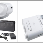
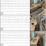
We present the case of an elderly male with OSA treated with CPAP, and in need of an ECG as apart of his care. The CPAP machine uses a hose and mask to deliver constant and steady air pressure (Figure 1). Three 12-lead ECGs were obtained with the CPAP machine on the bed (Figure 2, Panel A), touching the bed (Figure 2, Panel B) and off the bed (Figure 2, Panel C). From these ECGs, it is clear that electrical inference becomes less as the tubing attached to the CPAP machine is moved further away from the patient. It is important to note that the CPAP machine does not need to be in use or even turned on for this interference to occur (5). This type of interference could lead to the wrong diagnosis of cardiac arrhythmias (in this case the patient has real atrial flutter, but atrial and ventricular arrhythmias due to interference were previously described (5)); triggering the use of unnecessary medication with unpredictable results (5).
Figure 1. Parts of the Philips Respironics CPAP Machine (City, Country).
Figure 2. A: CPAP machine on the bed. Please note interference in the inferior leads.Baseline rhythm atrial flutter. B: CPAP machine touching the bed. Still significant interference can be seen. C: CPAP machine off the bed. Normalization of the ECG recording. Baseline rhythm atrial flutter.
CPAP machines are used in several OSA patients as the main treatment of choice. This device uses flexible tubing that connects the air outlet on the back of the device to the mask. The machine produces electrical oscillations that may hinder the procurement of an ECG and can generate artifacts that may interfere with the accurate diagnosis of the data attained.
The device works by adjusting humidity, oxygen supply, and pressure delivered, according to the therapy prescribed by the physician.
CPAP machines can produce interference on surface ECGs even if the contact between the patient and the machine is minimal (i.e. if the tubes are touching the bed). We are reporting this case to call to attention nurses and ECG technicians or other personnel obtaining ECG recordings. Interference may not be immediately obvious unless the ECG tech or RN performing the ECG is aware of how much a CPAP machine can interfere the recording. Completely disconnecting the CPAP machine from the wall and ensurin.
Conflict of interest
None
Acknowledgments
None
References
- Baranchuk, et al. It’s Time to Wake up!: Sleep Apnea and Cardiac Arrhythmias. OUP Academic, Oxford University Press 2008; 10.1093
- Digby, Genevieve C, and Adrian Baranchuk. Sleep Apnea and Atrial Fibrillation; 2012 Update. Current Cardiology Reviews 2012; 10.2174
- “CPAP Machines.” Philips Respironics, usa.philips.com/c-e/hs/sleep-apnea-therapy/sleep-apnea-machines.html.
- Baranchuk A, Pang H, Seaborn GEJ, Yazdan-Ashoori P, Redfearn DP, Simpson CS, Michael KA, Fitzpatrick M. Reverse Atrial Electrical Remodelling Induced by Continuous Positive Airway Pressure in Patients with Severe Obstructive Sleep Apnea. J Interventional Card Electrophysiol 2013; 36(3):247
- Suarez-Fuster, Laiden, Alexander B, Renaud R et al. Electrocardiographic Interference by a Sacral Neuromodulation Device. Journal of Electrocardiology 2017; 50(4):518-519
- Baranchuk A, Shaw C, Alanazi H, Campbell D, Bally K, Redfearn DP, Simpson CS, Abdollah H. ECG pitfalls and artifacts: The 10 Commandments. Crit Care Nurse 2009; 29:67-73
Copyright
© This work is licensed under a Creative Commons Attribution 4.0 International License. The images or other third party material in this article are included in the article’s Creative Commons license, unless indicated otherwise in the credit line; if the material is not included under the Creative Commons license, users will need to obtain permission from the license holder to reproduce the material. To view a copy of this license, visit http://creativecommons.org/licenses/by/4.0/


