J Med Discov (2019); 4(2):jmd19006; DOI:10.24262/jmd.4.2.19006;
Received February 19th, 2019,  Revised April 12nd, 2019,  Accepted April, 23rd, 2019,  Published May 08th, 2019.
Extremely elevated CA 19-9 levels in a patient with choledocholithiasis
Omar S. Mahmood1, Ali A. Mahdi1, Bashar Al hemyari2, Sahar E. Mohammadi2
1Sutter Institute for Medical Research, Sacramento, CA 95841
2University of California Riverside School of Medicine, GME Office, Riverside, CA 92501
* Correspondence: Omar S. Mahmood, Sutter Institute for Medical Research, Sacramento, CA 95841. Email: dr.omar86@gmail.com.
Abstract
On rare occasion, anorexia and weight loss could be the only presenting symptoms of choledocholithiasis mimicking a possible malignancy and warranting further evaluation. Although the specificity of a higher Carbohydrate Antigen 19-9 (CA 19-9) cut-off level (>1,000 U/mL) for pancreatic cancer is 99%, a high CA19-9 level of >1,000 U/mL often found in some benign diseases, however, the value is usually below 5000 U/ml. Cholestasis and Cholangitis seem to negatively affect the specificity of CA 19-9 in the evaluation of suspected pancreatic cancer and should always consider benign causes in the differential diagnosis even in the case of extremely high levels of CA 19-9 as simply managing these benign causes can result in a dramatic response in a relatively short period. Here we report a case that presented with weight loss, obstructive jaundice and extremely elevated CA 19-9 level due to choledocholithiasis.
Keywords: Choledocholithiasis, CA19-9, Pancreatic Cancer, Jaundice
Introduction
Although occasional patients with choledocholithiasis are asymptomatic, most patients are symptomatic. Symptoms include right upper quadrant or epigastric pain, nausea, vomiting, and obstructive jaundice1. On rare occasion, anorexia and weight loss could be the only presenting symptoms of choledocholithiasis mimicking a possible malignancy and warranting further evaluation2.
Carbohydrate Antigen 19-9 (CA 19-9) is a cell surface antigen that was first isolated by Koprowski in 1979 by utilizing immunologic tests (monoclonal Abs) on colonic cancer specimens 3,4 before its increasing value in the assessment of Pancreato-biliary diseases and neoplasms. With an upper limit of normal of 37 U/ml, the assay’s overall sensitivity is approximately 80%, and its specificity is 90%. Although the specificity of a higher CA19-9 cut-off level (>1,000 U/mL) for pancreatic cancer is 99%5, a high CA19-9 level of >1,000 U/mL often found in some benign diseases, such as common bile duct stones, acute cholangitis, acute pancreatitis, diabetes, and liver cirrhosis, but the value is usually below 5000 U/ml5-7.
Case Report
A 66-year-old male presented to the hospital with abdominal pain, malaise, and jaundice for one month duration. The pain was periumbilical, not radiated, dull, intermittent, no relieving factors, aggravated with food which resulted in food reluctance. He reported that his urine has been uncharacteristically dark over this period, but the stool had no real change in color or consistency. He had pruritus over his entire body. The patient endorsed constitutional complaints of intermittent fevers, chills, night sweats and overall fatigue with a headache, light-headedness but no focal weakness.
Additionally, he has lost 15.5 kg unintentionally and stated that he eats approximately 500 calories/day due to loss of appetite. The patient denied chest pain, dyspnoea, nausea, vomiting, diarrhea, constipation, melena or blurry vision. No recent travel or illness.
The patient had a non-contributory past medical and surgical history; Family history was significant for father with heart disease and mother with Leukaemia.
The patient quit smoking tobacco and drinking alcohol for eight years ago. He denied recreational or illicit drug use. He worked as a computer programmer. Currently Lives alone in an assisted living facility.
On general examination, the patient was alert, oriented and was in no acute distress. He had abnormal conjunctiva with icteric sclera and jaundice. Abdominal examination revealed a soft abdomen, mild deep epigastric tenderness, hepatomegaly, and active bowel sounds.
Investigations
Liver function test revealed a cholestatic pattern with elevated alkaline phosphatase (258 U/L), total bilirubin (27.1mg/dl) and mildly elevated transaminases, along with impaired liver synthetic functions demonstrated by prolonged Prothrombin time (18.8 seconds) and low albumin (2.2 g/dl).
Complete blood count revealed slightly elevated WBC (11.9 k/mm3) with neutrophil predominance on differential count (78.4%).
Renal function was normal.
CA 19-9 was markedly elevated (9272 U/ml).
Other investigations were negative for viral hepatitis serology and pancreatic lipase.
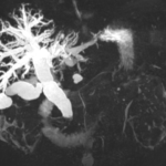
Figure 1. MRCP was significant for showing choledocholithiasis with at least two discrete gallstones in the distal CBD just proximal to the ampulla. The smaller more distal stone measured 8mm while the larger more proximal stone measured 1.5 cm.
Imaging Studies
Ultrasound of the gallbladder and liver showed a dilated common bile duct (CBD) measuring 22mm along with intrahepatic ductal dilatation measuring up to 10 mm Gallbladder wall thickness was normal, no peri-cholecystic fluid, no gallstones or sludge are visible inside the gallbladder.
CT scan of the abdomen with/without contrast revealed similar finding with no significant pancreatic lesion and was recommended MRCP/ERCP for further evaluation.
MRCP (Figure-1) was significant for showing choledocholithiasis with at least two discrete gallstones in the distal CBD just proximal to the ampulla. The smaller more distal stone measured 8mm while the larger more proximal stone measured 1.5 cm.
The visualized pancreas is unremarkable. There was no evidence of peri-pancreatic inflammation or pancreatitis.
Differential Diagnosis
Pancreatic Cancer
Cholangiocarcinoma
Common Bile duct stones
Chronic Pancreatitis
Ascending Cholangitis
Figure 2. ERCP demonstrated a bulging major papilla, moderate localized biliary stricture with severely dilated intrahepatic and extrahepatic duct and evidence of Cholangitis
Treatment
The patient was treated with IV fluids and parenteral antibiotics and scheduled for ERCP.
ERCP (Figure-2) demonstrated a bulging major papilla, moderate localized biliary stricture with severely dilated intrahepatic and extrahepatic duct and evidence of Cholangitis. A biliary sphincterotomy performed, and one temporary stent placed into the common bile duct. Brush biopsy of the CBD performed; the pathology report was negative for malignancy.
Endoscopic Ultrasound performed, and FNA of the common distal duct done, the pathology report was negative for malignancy.
Outcome and Follow up
The patient admitted for four days in the hospital, during this period his Bilirubin went down from 27mg/dl at admission to 7mg/dl at discharge.
AST from 96 U/L at admission to 41U/L at discharge.
ALT from 76 U/L at admission to 38 U/L at discharge.
ALP from 258 U/L at admission to 169 U/L at discharge.
CA 19-9 from 9272 U/ml at admission to 193 U/ml at discharge.
Outpatient follows up in 6 weeks
Bili 1.3, AST/ALT within normal limits, ALP 133.
The stent was removed.
Discussion
During our review for similar cases, Murohisa et al. presented a case of bile duct stone with cholangitis and high serum CA 19-9 level (60,000 U/ml), which returned to the normal six weeks later8.In another case reported by Marcouizos et al., a case of Choledocholithiasis with acute Cholangitis resulting in extremely high levels of CA 19-9 (99070 U/ml) which returned to normal levels within two months of the surgical removal of the stone9. A third case reported by Korkmaz et al., presenting a case of cholelithiasis, choledocholithiasis with acute cholangitis who had very high levels of CA 19-9 (9586 U/ml) that interestingly lowered to reach the level 50 U/ml within six days after successful biliary drainage.10
Another unique case was reported by Nasser et al. describing a morbidly obese patient presented with choledocholithiasis and high CA 19-9 levels (38310 U/ml) and two attempts of ERCP were done however his bile duct could not be cannulated due to anatomic distortion from his body habitus. Even though no definitive treatment achieved, CA 19-9 level done one week later showed a normal value of 36 U/ml11.
The pathophysiology behind the CA19-9 elevation in benign biliary disease is thought to arise from12:
- Excessive bile duct pressure stimulated bile duct cells to produce more CA19-9.
(II) Inflammation stimulated epithelial hyperplasia, leading to an increase in cells secreting CA19-9.
(III) Biliary obstruction and cholestasis decreased the clearance of CA19-9.
(IV) When the bile duct stones and inflammation subsided, the bile duct cells normalized, and CA19-9 also returned to normal levels.
In the study conducted by Kim et al. patients were divided into two groups then CA19-9 proved to be more useful for the group without cholangitis or cholestasis than for the group with cholangitis or cholestasis (p < 0.05). Therefore, the use of CA19-9 for the differentiation of pancreatic-biliary cancer should be applied individually, depending on the clinical situation 13.
In another study by Dogan et al. CA 19-9 levels were seen to be associated with biliary obstruction and cholangitis but not with the number and size of stones in patients with choledocholithiasis or cholangitis.14
Jalanko et al. reported that elevation of CA 19-9 levels found in 35% of 14 patients with choledocholithiasis; 10 of them (71%) had obstructive jaundice, but none had cholangitis. In that study, the highest CA 19-9 value was 440 U/ml.15
As a conclusion, cholestasis and cholangitis seems to negatively affect the specificity of CA 19-9 in the evaluation of suspected pancreatic cancer and should always consider benign causes in the differential diagnosis even in the case of extremely high levels of CA 19-9 as simply managing these benign causes can result in dramatic response in a relatively short period of time.
Conflict of interest
None
Acknowledgments
None
References
1. Mustafa A Arain, M. L. F., MD. in Uptodate (ed MD Shilpa Grover, MPH, AGAF) (Wolters Kluwer, 2017).
2. Lissoos, T. W., Hanan, I. M. & Blackstone, M. O. Anorexia and weight loss as the solitary symptoms of choledocholithiasis. South Med J 86, 239-241 (1993).
3. Koprowski, H. et al. Colorectal carcinoma antigens detected by hybridoma antibodies. Somatic Cell Genet 5, 957-971 (1979).
4. Koprowski, H., Herlyn, M., Steplewski, Z. & Sears, H. F. Specific antigen in serum of patients with colon carcinoma. Science 212, 53-55 (1981).
5. Steinberg, W. The clinical utility of the CA 19-9 tumor-associated antigen. Am J Gastroenterol 85, 350-355 (1990).
6. Mimidis, K., Anagnostoulis, S., Iakovidis, C. & Argyropoulou, P. Remarkably elevated serum levels of carbohydrate antigen 19-9 in cystic duct and common bile duct lithiasis. J Gastrointestin Liver Dis 17, 111-112 (2008).
7. Barone, D. et al. CA 19-9 assay in patients with extrahepatic cholestatic jaundice. Int J Biol Markers 3, 95-100 (1988).
8. Murohisa, T., Sugaya, H., Tetsuka, I., Suzuki, T. & Harada, T. A case of common bile duct stone with cholangitis presenting an extraordinarily high serum CA19-9 value. Intern Med 31, 516-520 (1992).
9. Marcouizos, G., Ignatiadou, E., Papanikolaou, G. E., Ziogas, D. & Fatouros, M. Highly elevated serum levels of CA 19-9 in choledocholithiasis: a case report. Cases J 2, 6662, doi:10.4076/1757-1626-2-6662 (2009).
10. Korkmaz, M., Unal, H., Selcuk, H. & Yilmaz, U. Extraordinarily elevated serum levels of CA 19-9 and rapid decrease after successful therapy: a case report and review of literature. Turk J Gastroenterol 21, 461-463 (2010).
11. Eiad Nasser, A. A., Dany Raad, Paula Burkard. Unusually Elevated CA 19-9
in Choledocholithiasis. Practical Gastroenterology, 52-54 (2013).
12. Sheen-Chen, S. M. et al. Extremely elevated CA19-9 in acute cholangitis. Dig Dis Sci 52, 3140-3142, doi:10.1007/s10620-006-9164-7 (2007).
13. Kim, H. J. et al. A new strategy for the application of CA19-9 in the differentiation of pancreaticobiliary cancer: analysis using a receiver operating characteristic curve. Am J Gastroenterol 94, 1941-1946, doi:10.1111/j.1572-0241.1999.01234.x (1999).
14. Dogan, U. B., Gumurdulu, Y., Golge, N. & Kara, B. Relationship of CA 19-9 with choledocholithiasis and cholangitis. Turk J Gastroenterol 22, 171-177 (2011).
15. alanko, H. et al. Comparison of a new tumour marker, CA 19-9, with alpha-fetoprotein and carcinoembryonic antigen in patients with upper gastrointestinal diseases. J Clin Pathol 37, 218-222 (1984).
Copyright
© This work is licensed under a Creative Commons Attribution 4.0 International License. The images or other third party material in this article are included in the article’s Creative Commons license, unless indicated otherwise in the credit line; if the material is not included under the Creative Commons license, users will need to obtain permission from the license holder to reproduce the material. To view a copy of this license, visit http://creativecommons.org/licenses/by/4.0/


