J Med Discov (2018); 3(4):jmd18043; DOI:10.24262/jmd.3.4.18043;
Received November 08th, 2018, Revised December 03rd, 2018, Accepted December 12nd, 2018, Published December 17th, 2018.
Chronic Ischemic Ulcer Myiasis
Ammar ELJack1,*, Mohamed Noureldin1, Khalid Mohamed1, Mohamedanwar Gandour1, Ahmed Subahi2
1Beaumont Hospital, Dearborn, Michigan, United States.
2Wayne State University, Detroit Medical Center, Detroit, Michigan, United States.
*Correspondence: Ammar ELJack, Email: ammar.eljack@beaumont.org.
Introduction
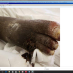
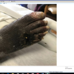
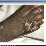
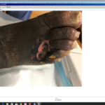
Wound Myiasis is a parasitic infestation of the wound by fly larvae; the female flies lay their eggs on the open wound which may take 8-24 hours to hatch.[1] Low socioeconomic status, poor hygiene, and exposed neglected wounds are the most common risk factors for acquiring myiasis.[1] Thus, the larval infestation is usually considered a complication of low-quality chronic ulcer care.[1] Wound Myiasis it is a rare infestation. However, human myiasis was reported in many primary care clinics in the United States.[2] We report the case of 61 years with past medical history hypertension, peripheral vascular disease, and end stage renal disease on hemodialysis who presented to ED with right foot pain and dorsolateral wound for two weeks before admission. The right foot wound was heavily infested with maggots with no evidence of local signs of secondary bacterial infections (figure 1); (Video 1). Magnetic resonance imaging (MRI) of the right foot showed osteopenia, and subacute fracture involving the proximal phalanx of the fourth toe but no osteomyelitis or abscess. Angiography of the right foot showed a poor flow to lower leg. The patient was treated with wound debridement, maggots’ removal, local wound care, and IV vacnomycin and zosyn for total of 7 days (figure 2). However, blood and wound cultures did not grow any organism. The wound healed uneventfully after this course of treatment. Management of chronic ulcers is usually challenging and needs multidisciplinary team collaborations. Therefore, over the last decade, after FDA approval in 2007 maggot debridement therapy (MDT) have been implemented as a novel biological approach in wound management.[3, 4] Maggot therapy can be used as a bridge to surgical debridement procedures, or as the primary debridement modality when surgical debridement cannot be performed.[5] MDT can also reduce the duration of antimicrobial therapy in some patients.[6] Three proposed actions are responsible for the therapeutic effects of MDT.
Fig 1. Showed 5x3 cm wound infected with maggots in dorsal-lateral aspect of the right foot (A) before wound examination; (B) after wound examination and removal of fibrotic exudate covering the wound.
First, maggots can secrete proteolytic enzymes that liquefy necrotic tissue, which is eventually get ingested by larvae.[3] Second, maggots have an antimicrobial action with enhanced bacterial biofilm eradication.[7] Finally, larvae can promote wound healing and stimulate granulation tissue growth.[8] Our patient’s circumstances of poor hygiene, neglect, and peripheral vascular disease substantially increased his risk of secondary bacterial wound infection. However, the presence of the associated larvae infestation had a natural wound debridement effect and helped to prevent bacterial infection.
Fig 2. Showed the patient’s right foot wound following surgical debridment (A) day one following the debridment ; (B) day two following the debridment.
Video 1: showed the patient’s right foot wound infected with maggots.
Conflict of interest
The authors have declared that no conflict of interest exists.
This case report did not receive any specific grant from funding agencies in the public, commercial, or not-for-profit sectors.
Patient consent obtained.
Acknowledgments
None
References
1. Francesconi F, Lupi O. Myiasis. Clin Microbiol Rev. 2012;25(1):79-105.
2. Huntington TE, Voigt DW, Higley LG. Not the usual suspects: human wound myiasis by phorids. J Med Entomol. 2008;45(1):157-9.
3. Andersen AS, Sandvang D, Schnorr KM et al. A novel approach to the antimicrobial activity of maggot debridement therapy. J Antimicrob Chemother. 2010;65(8):1646-54.
4. Paul AG, Ahmad NW, Lee HL et al. Maggot debridement therapy with Lucilia cuprina: a comparison with conventional debridement in diabetic foot ulcers. Int Wound J. 2009;6(1):39-46.
5. Nigam Y, Morgan C. Does maggot therapy promote wound healing? The clinical and cellular evidence. J Eur Acad Dermatol Venereol. 2016;30(5):776-82.
6. Armstrong DG, Salas P, Short B et al. Maggot therapy in “lower-extremity hospice” wound care: fewer amputations and more antibiotic-free days. J Am Podiatr Med Assoc. 2005;95(3):254-7.
7. Margolin L, Gialanella P. Assessment of the antimicrobial properties of maggots. Int Wound J. 2010;7(3):202-4.
8. Wollina U, Liebold K, Schmidt WD et al. Biosurgery supports granulation and debridement in chronic wounds–clinical data and remittance spectroscopy measurement. Int J Dermatol. 2002;41(10):635-9.
Copyright
© This work is licensed under a Creative Commons Attribution 4.0 International License. The images or other third party material in this article are included in the article’s Creative Commons license, unless indicated otherwise in the credit line; if the material is not included under the Creative Commons license, users will need to obtain permission from the license holder to reproduce the material. To view a copy of this license, visit http://creativecommons.org/licenses/by/4.0/


