J Med Discov (2018); 3(3):jmd17039; DOI:10.24262/jmd.3.3.17039;
Received August 26th,2018, Revised September 6th, 2018, Accepted September 7th, 2018, Published September 10th, 2018.
Mature Teratoma of the Bladder: A Rare Finding with a Possible Correlation to Teratomas elsewhere in the Body
Ahmed Oglah1,#, Samar Al-Rifaie1,#, Ali Allawi2, *
1Department of Urology, Baghdad medical city complex, Baghdad/Iraq, 10047
2Department of Nephrology, Medical city complex, Baghdad/Iraq, 10047
* Correspondence:Dr. Ali Allawi, chief of nephrology department at medical city complex, Baghdad, 10047, Iraq. E-mail: aliallawi15@yahoo.com. #Ahmed Oglah and Samar Al-Rifaie contributed equally to this work.
Abstract
Teratoma is extremely rare in the urinary bladder and can be found incidentally without any symptoms. Only a few cases were reported. We present a case of mature teratoma in the urinary bladder presenting as an asymptomatic bladder mass found incidentally during urinary tract imaging while looking for the cause of another condition, with a brief review of the literature.
Keywords: Bladder mass; Bladder, Teratoma, Benign
Introduction
Mature Teratomas are benign tumors; they consist of tissue from more than one germ cell layer (ectoderm, mesoderm and endoderm) and occur most commonly in the ovaries but may also be found at other sites. They are very rare in the urinary bladder. We present a case of mature teratoma in the urinary bladder of a 38-year old woman.
Case Report
A 38-year-old female referred to our Nephrology clinic for evaluation of left-sided hydronephrosis. She was first presented to the emergency room 1 month prior to her current visit due to acute severe left sided lower quadrant abdominal pain which was evaluated by an emergency Doppler ultrasound of the abdomen and pelvis that revealed a left ovarian mass with decreased blood flow suggesting an ovarian torsion. So a decision was made to do an emergency surgery. Intra-operatively the gynecologist found a left-sided ovarian mass with torsion, left oophorectomy was done and the specimen was sent to histopathology and found to be a mature ovarian teratoma. However, the follow-up ultrasound showed an incidental finding of left sided hydrouretronephrosis, the patient was referred to our Nephrology clinic for further evaluation.
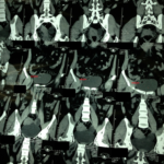
The patient didn’t report any irritative or obstructive urinary symptoms, ie; no dysuria, frequency, urgency, nocturia, hematuria, pilimiction or hesitancy and denies lower abdominal or flank pain. Examination was negative. The Investigation including urinalysis was normal. Abdominal and pelvic ultrasound revealed a left-sided hydroureteronephrosis without evidence of a stone. KUB x-ray was also negative. As such; a native CT scan of the abdomen and pelvis was ordered and showed a left sided hydroureteronephrosis with a cystic bladder mass with a central nidus of Calcification as shown in figure 1.
The specimen measured 4 × 3 × 2 cm and was grayish in appearance. Its cut surface showed a yellowish appearance with a tooth within it. The specimen was sent for histopathological exam and a mature teratoma of the bladder was confirmed by showing: hair follicles and sweat glands (Ectoderm) and a tooth (mesoderm).
Figure 1 : native CT scan of the abdomen showing a cystic bladder mass with central nidus of calcification ( Red arrow)
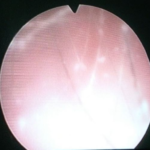
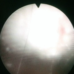
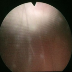
The patient was then referred to the Urology clinic, where they did a check cystoscopy. At cystoscopy, a single grey pedunculated mass measuring about 4 cm in diameter (Figures 2, 3 and 4) covered with hair was found arising just to the right of the midline of the bladder dome. The rest of the bladder mucosa was normal. The mass was excised using a transurethral resection of bladder tumor via a resectoscope and the specimen was sent for histological examination.
Figure 2: Check Cystoscopy image showing hairy grayish bladder mass.
Figure 3: Check Cystoscopy image showing hairy grayish bladder mass.
Figure 4: Check Cystoscopy image showing hairy grayish bladder mass.
Discussion
Teratomas presumably result from abnormal germ cells. A mature teratoma in the urinary bladder is an exceedingly rare ‘tumor'[1]’. We found only five cases reported and cited in the literature [2-6]. They usually contain hair and calcified material [4]. They may also be associated with bladder diverticula and vesical stones [3].On ultrasound imaging, a mature dermoid cyst appears as a thick-walled cystic mass with echogenic contents and calcification [7]. CT scan is the preferred diagnostic as it demonstrates fats with calcification [8]. The diagnosis of a mature cystic teratoma using CT imaging is straightforward because this modality is more sensitive for fat [9]. This tumor was a solitary tumor at the dome of the bladder. It contained calcified material. Histology confirmed skin, skin adnexal structures (sweat glands, hair follicles) and a tooth (mesoderm). This finding is important in that it enters the differential diagnosis of bladder mass, and the patient, as well as the surgeon, can be reassured since it is benign and will not need further treatment, unless it is symptomatic.
Conclusion
Although mature teratoma of the bladder is a rare incidence, it might coincidentally be found with teratomas of other organs, for example, with teratoma of the ovary. However, no evidence yet as of the association between the coincidental finding of multiple teratomas in the body. And no current recommendations to screen for teratomas elsewhere in the body after finding of an ovarian teratoma.
Conflict of interest
The authors declare that they have no competing interests.
Acknowledgments
Written consent was obtained from the patient prior to publication of this case report.
References
1.Eble JN, Young RH. Christofer DM Fletcher. 2nd ed. Churchill Livingston: China; 2000. Diagnostic Histopathology of tumours.
2.Cauffield EW. Dermoid cysts of the bladder. J Urol. 1956;75:801–804. [PubMed]
3.Lazebnik J, Kamhi D. A case of vesicalteratoma associated with vesical stones and diverticulum. J Urol. 1961;85:796–799. [PubMed]
4.Sabnis RB, Bradoo AM, Desai RM, Bhatt RM, Randive NU. Primary benign vesicalteratoma. A case report. Arch Esp Urol. 1993;46:444–445. [PubMed]
5.Misra S, Agarwal PK, Tandon RK, Wakhlu AK, Misra NC. Bladder teratoma: a case report and review of literature. Indian J Cancer. 1997;34:20–21.
6.Agrawal S, Khurana N, Mandhani A, Agrawal V, Jain M. Primary bladder dermoid: a case report and review of the literature. Urol Int. 2006;77(3):279-80.
7.Tandon A, Gulleria K, Gupta S, Goel S, Bhargava SK, Vaid NB. Mature ovarian dermoid cyst invading the urinary bladder. Ultrasound Obstet Gynecol. 2010;35(6):751-3.
8.Park SB, Kim JK, Kim KR, Cho KS. Imaging findings of complications and unusual manifestations of ovarian teratomas. Radiographics. 2008;28(4):969-83.
9.Schultheis J, Köhler F. Benign cystic teratoma (dermoid cyst) of the ovary perforated into the urinary bladder. Z UrolNephrol. 1977;70(10):733-7.
Copyright
© This work is licensed under a Creative Commons Attribution 4.0 International License. The images or other third party material in this article are included in the article’s Creative Commons license, unless indicated otherwise in the credit line; if the material is not included under the Creative Commons license, users will need to obtain permission from the license holder to reproduce the material. To view a copy of this license, visit http://creativecommons.org/licenses/by/4.0/


