J Med Discov (2018); 3(3):jmd18038; DOI:10.24262/jmd.3.3.18038;
Received August 26th, 2018, Revised September 18th, 2018, Accepted September 20th, 2018, Published September 26th, 2018.
Nephronophthisis in 11-year-old female presented with anemia
Ammar M. A. Algburi, M.D.*
Al-Yarmouk Teaching Hospital, Baghdad, Iraq.
* Correspondence: Ammar M. A. Algburi, Al-Yarmouk Teaching Hospital, Baghdad, Iraq. E-mail: dr.lami2020@gmail.com
Abstract
Here we report a case of nephronophthisis in an 11-year-old child with a known case of mental retardation, positive family history of renal failure, having pallor that is not responding to treatment with history of polyuria, polydipsia, and retinal hypoplasia.
Keywords: Nephronophthisis (NPHP); child; anemia; renal failure.
Introduction
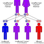
Nephronophthisis (NPHP) is a rare autosomal recessive cystic kidney disease (figure-1) and a leading genetic cause of renal failure in children and young adults that’s histologically characterized by interstitial fibrosis and corticomedullary cysts replacing normal renal tissue. The median age of NPHP induced renal failure in children is 13 years. The incidence of NPHP varies worldwide and ranges from 1 in 900,000 to 1 in 50,000. [1] The paucity of this disease and its presentation in mentally retarded female child prompted this case report.
Case Presentation
An 11-year-old female child admitted to our hospital as her mother stated that she was complaining from pallor for one-month duration. She is a known case of mental retardation presented with history of pallor for one-month duration of progressive onset that’s not associated with fever, blood loss, skin rash or change in color of urine. The family consulted a primary health care center where iron syrup was prescribed to her but her condition deteriorated by the development of poor feeding and decrease in her activity based on which the family consulted a private clinic where investigations revealed increase in renal function indices. Afterwards, her doctor referred her to our hospital where she was admitted to our nephrology unit because of history of polyuria. On admission, she had history of polyuria, about 11 time/day, started 5 months ago and associated with polydipsia and enuresis. There was no history of hematuria or dysuria. History and examination of other systems was unremarkable. In regards to vaccination, she completed her vaccinations since the age of 4 years according to her schedule and BCG scar is present at left forearm. However, she had history of developmental delay, sitting at age of one year, walking at age of 3.5 years, speaking one word at age of 2 years. speaking in 2 to 3 word-sentences at the age of 5 years, she got sphincter control at age of 8 years, she did not attend neither kindergarten nor school because of her mental retardation.
The past medical history revealed previous admission to the hospital for Transient Tachypnea of Newborn (TTN) in neonatal period and at age of 5 years, her family consulted an ophthalmologist who diagnosed her with bilateral retinal hypoplasia. Family history revealed that she has two cousins who are known cases of cerebral palsy and both of them died due to renal failure. On examination, her weight: 34 kg (on 25th centile), hight:135 cm (on 10th centile). Vital signs were all normal with normal blood pressure.
General examination revealed only mild leg edema. On Abdominal examination, there was no abdominal distention, organomegaly, palpable mass or shifting dullness. Neurological examination, she was alert with dysarthric speech, she had left eye movement limitation (left oculomotor apraxia), she had vertical nystagmus in all directions. On fundoscopy, there was pale retina. Other cranial nerves were intact. Tone was decreased in four limbs, power was +4 in upper and lower limbs bilaterally, reflexes were normal in upper and lower limbs bilaterally, sensory examination was normal, and impaired coordination in upper and lower limbs with ataxic gait.
Blood and urine investigations: Table. 1 Blood and urine indices.
| Blood index | value | Normal range |
| White blood cells (W.B.C) | 6.5 *10^9/l with normal differentiation | 4-10 x 10^9/L |
| Packed cell volume (P.C.V) | 24 % | 35-45% for females |
| Platelets (Plt) | 239*10^9/l | 150-400 x 10^9/L |
| Serum sodium (s.Na) | 132 mmol/l | 135-145 mmol/L |
| Serum potassium (s.K) | 4.3 mmol/l | 3.5-5 mmol/L |
| Serum calcium (s.Ca) | 0.7 mmol/l | 2-2.6 mmol/L |
| Serum phosphate (s.PO4) | 2.6 mmol/l | 0.8-1.5 mmol/L |
| Random blood sugar | 4.6 mmol/l | 3.9-5.6 mmol/L |
| Blood urea nitrogen | 27 mmol/l | 2.9-7.1 mmol/L |
| Serum creatinine | 356 μmol/L | 61.9-115 μmol/L |
| Prothrombin time (PT) | 13 seconds | 11-14 sec |
| Partial thromboplastin time (PTT) | 29.8 seconds | 20-40 sec |
| General Urine Examination | ||
| Specific gravity | 1000 | 1001-1035 |
| W.B.C. | 4-6/hpf | ≤2-5 WBCs/hpf |
| R.B.C | nill | ≤2 RBCs/hpf |
| Albumin | nill | nill |
| Blood film | Normochromic normocytic anemia with normal W.B.C. and Plt. |
Table. 2 Genetics defects underlying NPHP, associated features and other clinical phenotypes [3]
| Gene (protein) | Chromosome | NPHP type | Clinical features associated with NPHP | Other clinical phenotypes |
| NPHP1 (nephrocystin-1) | 2q13 | NPHP | Mild JS, mild RP, Cogan | JS |
| NPHP2/INVS (inversin) | 9q31 | Infantile NPHP | RP, liver fibrosis, situs inversus, hypertension, VSD | |
| NPHP3 (nephrocystin-3) | 3q22 | NPHP, Infantile NPHP | Liver fibrosis, RP, situs inversus | MKS |
| NPHP4 (nephrocystin-4 or nephroretinin) | 1p36 | NPHP | Cogan, RP | |
| NPHP5/IQCB1(nephrocystin-5) | 3q21 | NPHP | Severe RP | |
| NPHP6/CEP290(nephrocystin-6/CEP290) | 12q21 | NPHP | JS, severe RP | Isolated RP, JS, MKS, BBS |
| NPHP7/GLIS2 (GLIS2) | 16p | NPHP | ||
| NPHP8/RPGRIP1L(RPGRIP1L) | 16q | NPHP | JS | JS, MKS |
| NPHP9/NEK8 (NEK8) | 17q11 | NPHP, Infantile NPHP | ||
| AHI1 (Jouberin/AHI1) | 6q23 | NPHP | JS, RP | JS |
JS, Joubert syndrome type B; RP, retinitis pigmentosa; Cogan, oculomotor apraxia type Cogan; MKS, Meckel–Gruber syndrome; VSD, ventricular septal defect
Fig.1 genetic inheritance of Nephronophthisis
|
Eye/retinal disease Isolated oculomotor apraxia Retinitis pigmentosa Coloboma Nystagmus Ptosis Neurological disease Learning difficulties Cerebellar ataxia with vermis hypoplasia Hypopituitarism Encephalocoele Liver disease Elevation of hepatic enzymes Fibrosis, biliary duct proliferation Skeletal Phalangeal cone-shaped epiphyses Short ribs Postaxial polydactyly Skeletal dysplasia Others Situs inversus Cardiac malformations Bronchiectasis Situs inversus Cardiac malformations Bronchiectasis |
Table.3 Extra renal manifestations associated with NPHP [3]
Imaging Investigation
Chest X-Ray:
normal
Abdominal Ultrasound:
right kidney length 9.3 cm
Left kidney length 9 cm
Increase in echogenicity with ill-defined corticomedullary junction
Picture suggestive of parenchymal renal disease.
Abdominal CT scan:
Right kidney shoe normal size and position of 9.2 cm
Left kidney shows normal size of and position 9.1 cm
Both kidneys show no evidence of cysts or calcification
Differential diagnosis:
Neurogenic bladder lead to CRF.
Nephronophthisis.
Polycystic kidney disease.
Diagnosis and treatment: The patient was diagnosed with nephronophthisis and was given the followi2ng treatment:
Hemodialysis
Folic acid tab 5 mg/day
Vitamin D3 1000 IU/day
Calcium carbonate tab 500 mg/day
Ferrous syrup 40 mg * 2/day
Epirax (erythropoietin) vial 400 IU/wk
Discussion
Nephronophthisis (NPHP) is a cause of autosomal recessive cystic kidney disease as well as renal fibrosis and end-stage kidney failure that typically affects young adults and children.[2] Many of the genes have been described as the cause of this hereditary disease and many of which are expressed on the in centromeres and primary cilia, rendering he disease to be as ciliopathy disease (table-2). [3] The histopathological features of the disease consist of corticomedullary cysts, atrophy, and interstitial fibrosis that reflect clinically as reduced GFR and loss of tubular function in the range of polyuria, polydipsia, secondary enuresis and growth retardation in children and eventually end stage renal disease ESRD. [4] The widely used classification of the disease is based upon age of ESRD onset as infantile, that causes ESRD around the age of 1; juvenile and adolescent that cause ESRD around the ages of 13 and 19 respectively. The Juvenile form is the classical one which is characterized by polyuria and polydipsia symptoms and often anemia in patients within the first decade of life. Progressive loss of kidney function leads to ESRD at a median age of 13 years while other forms are rare. [5]
NPHP is characterized by inability to concentrate urine early in life that is responsible for polyuria and polydipsia. The disease may be easily misdiagnosed, as there is typically no severe hypertension, very little or no proteinuria. Besides extensive investigations of renal function, clinical phenotyping should also encompass a full neurological screening to assess for cerebellar signs and fundoscopy to assess for retinal degeneration.
A formal ophthalmological examination is advised. The role of renal biopsy in diagnosing NPHP should be limited to cases that are hard to be differentiated from other diseases. In most cases, a histopathological diagnosis should be preceded by a molecular genetic diagnostic approach, because genetic screening allows for early diagnosis and prevents complications of renal biopsy. NPHP1 mutations and deletions are the most frequent genetic cause of NPHP and may be screened for using standard PCR assays. [6]
NPHP should be considered in any child who presents with the following characteristics:
-Polyuria due to decreased concentrating ability with a relatively normal urinalysis and reduced urinary sodium conservation
-Progressive chronic kidney disease with a normal blood pressure
-Ultrasonography demonstrates normal or slightly decreased in size kidneys for age with increased echogenicity with loss of corticomedullary junction. There is no dilation of the urinary tract, and renal cysts are not typically identified on the initial ultrasound examination but may appear at a late stage of the disease. The diagnosis is also supported by the presence of associated extrarenal manifestations (table-3).
Various theories behind the pathogenesis of the NPHP have been described. The very early hypotheses stated that this disease was caused by some unknown nephrotoxic agent or an enzyme defect. [7] The finding of tubular basement membrane thickening led to a basement membrane hypothesis for the pathogenesis of NPHP. Later, the hypothesis that described NPHP as a ciliopathy emerged. [8] This hypothesis is strongly supported by multiple gene discoveries in NPHP with many of which are coding for the components of the cilia. A study that links between NPHP and cilia found that INVS mutations cause infantile NPHP and that the encoded protein inversin interacts with nephrocystin-1 and β-tubulin, co-localizing with them to the primary cilia of renal tubular cells. [9]
The treatment of NPHP is mainly supportive. Control of blood pressure and correction of water and electrolyte imbalances are priorities in affected children and young adults. Management of children in the early stage of the disease without renal impairment is focused upon replacing their ongoing water needs to prevent episodes of hypovolemia. Patients with NPHP uniformly progress to ESRD. Renal transplantation is the preferred therapy in patients with ESRD. However, management of complications arising from progressive renal failure such as anemia, symptoms of uremia and fluid overload are important alongside preparation for future renal replacement therapy.
In conclusion, we diagnosed this patient as having NPHP disease given the positive family history of renal failure, history of pallor that is not responding to iron treatment alone, history of polyuria, polydipsia and retinal hypoplasia, in addition to imaging and blood tests results. The patient had inability to conserve sodium because of defect of tubules leading to polyuria and polydipsia. The anemia is attributed to a deficiency of erythropoietin production by failing kidneys. She had no hypertension as nephronophthisis is a salt-losing nephropathy.
Conflict of Interest
None
Acknowledgments
Author discloses no acknowledgement. No funding was received for this case report.
References
1. Simms RJ, Hynes AM, Eley L, Sayer JA. Nephronophthisis: a genetically diverse ciliopathy. Int J Nephrol. 2011;2011:527137.
2. Delous M, Baala L, Salomon R, Laclef C, Vierkotten J, Tory K, et al. The ciliary gene RPGRIP1L is mutated in cerebello-oculo-renal syndrome (Joubert syndrome type B) and Meckel syndrome. Nat Genet. 2007;39(7):875-81.
3. Simms RJ, Eley L, Sayer JA. Nephronophthisis. Eur J Hum Genet. 2009;17(4):406-16.
4. Krishnan R, Eley L, Sayer JA. Urinary concentration defects and mechanisms underlying nephronophthisis. Kidney Blood Press Res. 2008;31(3):152-62.
5. Hildebrandt F, Strahm B, Nothwang HG, Gretz N, Schnieders B, Singh-Sawhney I, et al. Molecular genetic identification of families with juvenile nephronophthisis type 1: rate of progression to renal failure. APN Study Group. Arbeitsgemeinschaft fur Padiatrische Nephrologie. Kidney Int. 1997;51(1):261-9.
6. Otto EA, Helou J, Allen SJ, O’Toole JF, Wise EL, Ashraf S, et al. Mutation analysis in nephronophthisis using a combined approach of homozygosity mapping, CEL I endonuclease cleavage, and direct sequencing. Hum Mutat. 2008;29(3):418-26.
7. Waldherr R, Lennert T, Weber HP, Fodisch HJ, Scharer K. The nephronophthisis complex. A clinicopathologic study in children. Virchows Arch A Pathol Anat Histol. 1982;394(3):235-54.
8. Watnick T, Germino G. From cilia to cyst. Nat Genet. 34. United States2003. p. 355-6.
9. Otto EA, Schermer B, Obara T, O’Toole JF, Hiller KS, Mueller AM, et al. Mutations in INVS encoding inversin cause nephronophthisis type 2, linking renal cystic disease to the function of primary cilia and left-right axis determination. Nat Genet. 2003;34(4):413-20.
Copyright
© This work is licensed under a Creative Commons Attribution 4.0 International License. The images or other third party material in this article are included in the article’s Creative Commons license, unless indicated otherwise in the credit line; if the material is not included under the Creative Commons license, users will need to obtain permission from the license holder to reproduce the material. To view a copy of this license, visit http://creativecommons.org/licenses/by/4.0/


