J Sci Discov (2017); 1(1):jsd17007; DOI:10.24262/jsd.1.1.17007;
Received May 23rd, 2017, Accepted June 3rd, 2017, Published June 5th, 2017
Pseudomelanosis Duodeni: An Interesting and Rare Finding At Upper G.I. Endoscopy
Aws Alameri1,#, Asseel Albayati1,#, Nisar Ahmed1,*
1Department of Gastroenterology, Park Plaza Hospital, Houston , TX, 77004
* Correspondence: Dr. Nisar Ahmed, M.D. F.A.C.G. , Chief of Gastroenterology, Park Plaza Hospital, Houston, TX 77004, USA. Email: nisarahmedmd@yahoo.com.
# Aws Alameri and Asseel Albayati contributed equally to this work.
Abstract
Pseudomelanosis Duodeni is an incidental endoscopic finding that is seen as splotches of brown-black pigment of the duodenal mucosa. Only a few cases have been reported. Sometimes it was found to be associated with certain medications and systemic diseases. It is a benign condition and does not seem to have clinical consequences but needs to be recognized to distinguish it from more serious conditions like Malignant Melanoma.
Keywords: Pseudomelanosis Duodeni; Duodenum; Pigment; Benign
Introduction
Pseudomelanosis Duodeni is a rare condition of the Gastrointestinal tract that is seen as spotty brown to black pigmentation of the duodenal mucosa during endoscopy [1], [2]. It raises the possibility of malignant melanoma, but a simple biopsy helps to rule it out.
Case Report
A 71 years old African American male patient presented to us complaining of occasional difficulty in swallowing and a very bitter taste in his mouth. His symptoms started 2 months ago, and were getting worse despite of medications. He gave history of Diabetes Mellitus type 2, and Hypertension, also he has a family history of Diabetes mellitus type 2. He was mildly anemic; with Hb 11.3 g/dl, but he denied use of any iron supplements, and gave no history of hematemesis, melena, or blood transfusion.
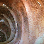
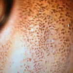
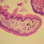
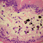
Upon endoscopy, melanosis-like mucosal pigmentations were seen in the duodenum (Fig. 1 & 2). Biopsies taken from these pigmentations are shown in (Fig. 3 & 4).
Figure 1. Duodenum, spotty mucosal pigmentation.
Figure 2. Duodenum, close view.
Figure 3. Histopathological specimen of duodenal mucosa of Pseudomelanosis Duodeni.
Figure 4. Histopathological specimen of duodenal mucosa, brown-pigmented deposition in the Lamina propria of duodenal villus.
Discussion
Pseudomelanosis Duodeni was first described in 1976 as Melanosis Duodeni, but was renamed Pseudo melanosis as the pigment is not composed of melanin but rather primarily of Iron (positive Prussian blue stain) , and Sulfate. Other chemicals like lipomelanin, calcium, potassium, aluminum, magnesium and silver can also be detected [1], [2]. The diagnosis is usually incidentally made by endoscopy and biopsy which usually show a peculiar brown pigment in the Macrophage in Lamina Propria of Duodenal Villi [3]. It is also worthy to mention that the histiocytes in Pseudomelanosis Duodeni are positive for CD168 and CD68. The clinical significance highlights the importance to exclude other causes like metastatic melanoma, hemochromatosis, and brown bowel syndrome. Moreover, it was found that Pseudomelanosis Duodeni is a different entity from Melanosis Coli, and unlike the latter is not related to laxative use. [4].
There has been few similar cases reported, and it was shown that Pseudomelanosis Duodeni is more predominant in females in their sixties and seventies. Although correlations with drug use (Charcoal, hydralazine, furosemide, hydrochlorothiazide, propranolol, and iron supplements) and systemic diseases (hypertension, chronic renal disease, gastric hemorrhage, and diabetes mellitus) have been noted, the etiology of Pseudomelanosis Duodeni has not been yet elucidated [5], [6], [7].
Conclusion
Pseudomelanosis Duodeni, so far, is a benign condition and carries no significant consequences. However, this does not mean to overlook this finding, it is important to exclude serious conditions especially metastatic melanoma.
Competing interests
The authors declare that they have no competing interests.
Acknowledgments
None
References
1. Bisordi WM, Kleinman MS. Melanosis duodeni. Gastrointest Endosc.1976;23:37–38.
2. Banai J, Fenyvesi A, Gonda G, Petö I. Melanosis jejuni. Gastrointest Endosc. 1997;45:432–434.
3. Giusto D, Jakate S. Pseudomelanosis duodeni: associated with multiple clinical conditions and unpredictable iron stainability – a case series. Endoscopy 2008; 40: 165-167.
4. Abumoawad A, Venu M, Huang L, Ding XZ. Pseudomelanosis duodeni: a short review. Am J Digest Dis 2015;2(1):41-45.
5. Cantu JA, Adler DG. Pseudomelanosis duodeni. Endoscopy. 2005;37:789.
6. eL-Newihi HM, Lynch CA, Mihas AA. Case reports: pseudomelanosis duodeni:association with systemic hypertension. Am J Med Sci.1995;310:111–114.
7. Ghadially FN, Walley VM. Pigments of the gastrointestinal tract: a comparison of light microscopic and electron microscopic findings. Ultrastruct Pathol. 1995;19:213–219.
Copyright
© This work is licensed under a Creative Commons Attribution 4.0 International License. The images or other third party material in this article are included in the article’s Creative Commons license, unless indicated otherwise in the credit line; if the material is not included under the Creative Commons license, users will need to obtain permission from the license holder to reproduce the material. To view a copy of this license, visit http://creativecommons.org/licenses/by/4.0/


