Journal of Medical Discovery (2016); 1(1): jmd16002; Received September 30th, 2016, Revised December 27th, 2016, Accepted January 2nd, 2017,
Published February 2nd, 2017
Schwannoma of recto-sigmoid colon: a rare occurrence
Zubair Shahid Bashir1, Tesingin Destiny Uwawah1, Nisar Ahmed1
1 Department of Gastroenterology, Park Plaza Hospital, Texas Medical Center, Houston, Texas.
* Correspondence: Zubair Shahid Bashir, Department of Gastroenterology, Park Plaza Hospital, Texas Medical Center, Houston, Texas.
Email: shahid.zubair08@gmail.com. Tel: +1 708-340-2832. Postal Address: 2386-B Birch Run Circle, Herndon, VA, 20171
Abstract
Schwannomas are usually slow growing, benign and asymptomatic tumors. These arise from Schwann cells. The Schwann cells insulate the nerves which allow rapid transmission of impulses between the nodes of Ranvier. Schwannomas are commonly found in the antrum of stomach constituting 6.3% of the gastric mesenchymal tumors.
We present a case of incidental schwannoma in the recto-sigmoid colon. Though these are mostly asymptomatic but do have a tendency to cause GI bleed, obstruction, ulceration, abdominal pain and undergo malignant transformation (1).
Keywords: Schwannoma, mesenchymal tumors, colon, GI bleed and malignant transformation
Case Report
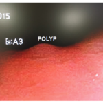
A 51year old African American female patient underwent screening colonoscopy. She had family history of colon cancer in father and colon polyps in mother. She is known to have schizoaffective disorder, treated with lorazepam and duloxetine. She specifically had history of intermittent constipation for couple of years. On colonoscopy a 5mm sessile polyp was identified in recto-sigmoid colon (Fig. 1). It was removed with hot biopsy cautery. The tissue was sent to laboratory for examination. Biopsy result was consistent with Schwannoma.
Figure 1. Gross picture of polyp.
Discussion
Schwannomas are homogenous tumors of Schwann cells. These cells insulate the peripheral nerves and allow rapid transmission of impulses between CNS and periphery. They produce symptoms by impinging on the nerves. Mostly schwannoma occur in peripheral nerves of head and neck region. Rarely these are seen in the GIT.
The GIT schwannomas have a tendency to ulcerate, bleed, enlarge and cause obstruction. The chance of malignant transformation is 0.3% (2). These tumors also exist with other extra and intra-intestinal tumors; primary adeno-carcinoma of ascending colon (3). An association with Hodgkin Lymphoma and Von Recklinghausen disease was reported by Qasi et al. (2). Despite occurring in the submucosal layer of GI tract they can enlarge up to 120mm in size and cause obstruction (4).
The exact etiology of schwannomas is not known. We hypothesize that chronic irritation of GI lining which leads to stimulation of Schwann cell over growth can contribute to the formation of schwannoma since the patient had history of several years of constipation. The continuous irritation over years with periods of intermittent constipation may lead to growth of normal Schwann cell layer.
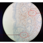
This irritation is caused by eating hard food like nuts, hard-dried fecal matter and further aggravated by straining to pass feces. The other common tumors of GI submucosa such as GIST, GNAT, leiomyoma and leiomyosarcoma can be differentiated by microscopy and immunohistochemistry. Schwannomas consist of densely packed spindle cells called Antoni A area (Verocay bodies) and loosely packed spindle cells called Antoni B area in myxoid stroma (Fig. 2).
Figure 2. Microscopic picture of Gastro-intestinal Schwannoma
Moreover, on immune-histochemistry schwannomas express S100 and vimentin protein unlike other tumors. These do not express the CD 117 antigen and are usually negative for CD 34 also. This is not the case with GISTs. Schwannomas are also negative for SMA, in contrast to leiomyomas. GANTs on the other hand, are usually negative for S-100 protein and GFAP and most are positive for CD 117 and CD 34 (5).
Competing interests
The authors declare that they have no competing interests.
Acknowledgments
There are no financial disclosures to be made.
References
1. S Atmatzidis K. Gastric schwannoma: a case report and literature review. Hippokratia. 2012;16(3):280. Available at: http://www.ncbi.nlm.nih.gov/pmc/articles/PMC3738740/. Accessed April 21, 2016.
2. Qasi Sabir E. Schwannoma of the rectum: A case report. Saudi Journal of Gastroenterology. 1996;2(3):164. Available at: http://www.saudijgastro.com/article.asp?issn=1319-3767;year=1996;volume=2;issue=3;spage=164;epage=167;aulast=Qasi. Accessed April 21, 2016.
3. Vasilakaki T, Skafida E, Arkoumani E et al. Synchronous Primary Adenocarcinoma and Ancient Schwannoma in the Colon: An Unusual Case Report. Case Reports in Oncology. 2012;5(1):164-168. doi:10.1159/000337689.
4. Hou YY e. Schwannoma of the gastrointestinal tract: a clinicopathological, immunohistochemical and ultrastructural study of 33 cases. – PubMed – NCBI. Ncbinlmnihgov. 2016. Available at: http://www.ncbi.nlm.nih.gov/pubmed/16623779/. Accessed April 21, 2016.
5. Atmatzidis S, Chatzimavroudis G, Dragoumis D, Tsiaousis P, Patsas A, Atmatzidis K. Gastric Schwannoma: A case report and literature review. Hippokratia. 2012;16(3):280-2. Available at: https://www.researchgate.net/publication/255736515_Gastric_Schwannoma_A_case_report_and_literature_review. Accessed April 21, 2016
Copyright
© This work is licensed under a Creative Commons Attribution 4.0 International License. The images or other third party material in this article are included in the article’s Creative Commons license, unless indicated otherwise in the credit line; if the material is not included under the Creative Commons license, users will need to obtain permission from the license holder to reproduce the material. To view a copy of this license, visit http://creativecommons.org/licenses/by/4.0/


