J Med Discov (2018); 4(1):jmd18046; DOI:10.24262/jmd.4.1.18046;
Received November 21st, 2018, Revised December 14th, 2018, Accepted December 18th, 2018, Published December 28th, 2018
Palpitations in patient with Wolff Parkinson White syndrome: a case report
Badie Batti¹, Ehab Embaby²
¹University of Baghdad, Internal medicine department, Baghdad, Iraq 10071, Email: Badiesabah@yahoo.com.
²Ehab Embaby M.B.B.Ch., Ain shams University hospital, Cairo, Egypt 11566, Email: Ehab.embaby1992@gmail.com
* Correspondence: Badie Batti M.B.Ch.B, University of Baghdad, Internal medicine department, Baghdad, Iraq 10071, Email: Badiesabah@yahoo.com.
Abstract
Palpitations are a common presenting symptom in the emergency department (ED). Typically, the cause of palpitations is benign, especially in otherwise young, healthy patients. However, physicians must be aware of more serious underlying conditions. Among patients with Wolff Parkinson White (WPW) syndrome, one-third were estimated to have atrial fibrillation (AF) at presentation. In this case, we present a patient with recurrent episodes of palpitations and highlight the diagnostic challenge.
Keywords: Palpitations, Wolff Parkinson White Syndrome, Atrial fibrillation.
Introduction
The normal conduction system of the heart confines movement of electrical impulses from the atria to the ventricles to a single common pathway through the Atrioventricular (AV) node [1].
Wolff-Parkinson-White (WPW) syndrome is a conduction disturbance in which atrial impulses are transmitted to the ventricles by an accessory pathway besides the normal AV conduction pathway.
Patients with this syndrome may present with palpitations, syncope [2], and/or sudden death. Multiple Patterns are noted among 0.1 to 0.5% of the population [3]. The pattern is dependent not only on the location of accessory pathway, but also on the properties of (AV) node and His-Purkinje system.
Spontaneous normalization of ECG with intermittent preexcitation is reported in 20% to 30% of WPW; dynamic QRS variations in WPW syndrome were noted several years ago [4].
We report a case of a young lady complaining of episodic palpitations associated with shortness of breath and anxiety, she was diagnosed with panic disorder after a negative initial emergency department workup and outpatient assessment with her primary care physician and cardiologist.
Surprisingly she had a recurrent episode of palpitation in which she was admitted back to the Emergency department for tachycardia with irregular rhythm proven to be AF which was treated accordingly to reveal a baseline ECG changes consistent with WPW syndrome.
Case Presentation
A 36-years-old female presented to ED complaining of palpitations described as racing of her heart, started 30 minutes prior to her arrival when she was taking a shower associated with shortness of breath and lightheadedness.
She reported feeling exhausted of her energy, being afraid of losing control of herself and having concerns about recurrent episodes of chest symptoms, but she denied any chest pain, orthopnea, exertional dyspnea, shaking movements, changes in vision or hearing, loss of consciousness, weakness or numbness.
Review of other systems was insignificant; However, review of past medical history revealed 3 previous episodes of similar chest complaints for the past 4 years with no aggravating or relieving factors terminating spontaneously and a previous ED admission with a negative ECG and routine blood tests included (Complete blood count and basic metabolic panel). She was subsequently diagnosed with Panic disorder and prescribed Fluoxetine (Prozac) 20 mg tablets after a negative cardiac workup that included Exercise tolerance test (ETT), 24-hr Holter monitoring and Echocardiography in which she reported no symptoms while doing.
She also stated that she had no follow up with her primary care physician for the last year until her current episode.
Review of her family history was insignificant for sudden death or major illness; However social and dietary history revealed that she used to exercise 3-4 times weekly, but no use of caffeine beverages or illicit drugs.
On Physical exam, our patient is a young well-built female looked in acute distress, she was alert and oriented to time, place and person.
Her vitals were: HR: 220 b/min irregular rhythm, BP: 90/60 mmHg, Temp: 37.3C° (99.14F°), RR: 18 breaths/min, O2 saturation 99% on 2 L.
She had facial and conjunctival pallor with sweating; Neck exam revealed no goiter; Heart exam revealed S1, S2 with no murmurs, rubs and gallops; Lungs were clear to auscultation with no added sounds; Extremities were cool on touch with a radial pulse 1+ bilaterally but no tremors.
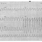
Upon physical exam, ECG was ordered to reveal irregular wide complex tachycardia a t a rate of 230 b/min in Fig. (1)
Fig 1. A 12-lead electrocardiogram showing a heart rate of 230 bpm (23 QRS in 30 large squares * 10 seconds) and irregular wide QRS complex tachycardia without P wave consistent with AF with rapid ventricular response.
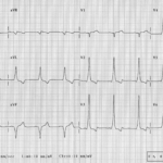
Because of dynamic instability, the Adult Cardiac Life Support (ACLS) algorithm was followed with a synchronized cardioversion, the patient converts into a sinus rhythm successfully and a repeated ECG showed shortened PR interval and slurred initial portion of QRS complex (known as delta wave) in Fig. (2) in which she was admitted to cardiac care unit and prescribed (Procainamide) infusion.
The patient was subsequently referred to an electrophysiologist for radiofrequency ablation of the accessory conduction pathway to prevent life threatening cardiac arrhythmias.
Discussion
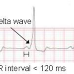
The diagnosis of WPW syndrome with AF was made on the basis of the patient’s history in conjunction with the classic ECG findings: short PR interval (normal is 3-5 small squares), slurred initial portion of QRS “the delta wave” Fig. (3), and a wide QRS complex changes (normal is less than 3 small squares).
Fig 2. A postconversion electrocardiogram showing sinus rhythm with shortened PR interval (prominent in V2-V5) and slurring of the wide QRS complex (I , aVL, V2-V4).
The arrhythmia being irregularly irregular; this is an important fact in recognizing atrial fibrillation and the history of previously undiagnosed
paroxysms of palpitations and shortness of breath is common in cases of supraventricular tachycardia.
The subtle findings on the baseline ECG are often overlooked, and young patients can be diagnosed with other disorders, such as anxiety or panic attacks.
The wide complex tachycardia represents activation of the ventricles through a pathway outside of the normal conduction system [5-7], this is known as preexcitation and the heart rates of 170-300 b/min are consistent with the cycle of atrial fibrillation, with almost 1:1 activation of the ventricles through this accessory pathway.
Since these rapid heart rates lack the normal delay in conduction (an intrinsic protective mechanism) from the atrioventricular node [8], the ventricular rhythm can degrade into Ventricular fibrillation, resulting in sudden cardiac death [9].
This is a life-threatening event and requires immediate intervention, even if the patient appears hemodynamically stable. In this case, the ED staff made use of the ACLS algorithms because of the patient’s hemodynamic instability.
Synchronized cardioversion is a reasonable option for treatment of this rhythm. Alternatively, if the patient is a bit more stable, a bolus of (amiodarone), which is also part of the ACLS algorithm, could selectively decrease conduction through the bypass tract relative to the atrioventricular node, resulting in a break in the rhythm [10]; However there are some reports of ventricular fibrillation following administration [11].
If the rhythm is recognized immediately, (procainamide) is a more effective option for stopping this arrhythmia [12].
The bundle of Kent, is a communicating tract between the left atrial appendage and left ventricle, is a classic example of a preexcitation accessory pathway and localization of the various types of accessory pathways, as in table (1) [13] can often be achieved through the baseline ECG.
Atrioventricular accessory pathways are not the only mechanism of early ventricular activation. There are rare reports of atrio-hisian pathways, a condition known as Lown-Ganong-Levine (LGL) syndrome [14]. In LGL syndrome, the ECG demonstrates a short P-R interval (usually < 0.12 sec) without a delta wave and with a normal QRS complex.
Fig 3. Initial slurred wave prior to QRS complex on electrocardiograph is a delta wave.
In many cases of narrow-complex tachycardia, Adenosine can be helpful in both the diagnosis and, depending on the underlying arrhythmia, the treatment to stop the cycle of the arrhythmia; however, patients who have the typical narrow-complex atrioventricular reentrant tachycardia associated with WPW Syndrome can theoretically be at risk for harm. This is because Adenosine can prolong conduction and refractory time in the atrioventricular node, promoting conduction down the accessory pathway, resulting in arrhythmias [15-16].
Digoxin, beta blockers and non-dihydropyridine calcium channel blockers are contraindicated in preexcitation tachycardia because they may shorten the refractory period and enhance conduction over the bypass tract, resulting in even faster conduction to the ventricles and increasing the risk for ventricular fibrillation.
Some patients with WPW syndrome are at risk for sudden death and in these patients, a cardiac electrophysiology study and radiofrequency catheter ablation may be definitive and curative.
Table 1: Localization of the bypass tract from analysis of delta wave axis and polarity of QRS complexes on ECG.
| Pathway site | Polarity of main QRS complex | QRS axis | Delta axis | ||
| Lead V1 | Lead V2 | Lead V3 | |||
| Anteroseptal | Negative | Negative | Negative | Normal | Normal |
| Right lateral | Negative | Negative | Negative | Left | Left |
| Right posteroseptal | Negative | Positive | Positive | Left | Left |
| Left posteroseptal | Positive | Positive | Positive | Left | Left |
| Left lateral | Positive | Positive | Positive | Inferior | Inferior |
Conclusion
This case highlights the importance of follow up and suggests insensitive diagnostic modalities including 24-hr Holter monitoring to identify all types of AF which can be the only presenting sign of underlying cardiac conduction system disturbance.
When confronted with an irregular wide QRS tachycardia, physicians should always be concerned that this might represent preexcited tachycardia as early recognition and treatment allows rapid restoration of sinus rhythm and may decrease morbidity and mortality.
Conflict of interest
None
Acknowledgments
We would like to thank the Internal medicine department in Alkindi teaching hospital for their help and support.
References
1. Marillo CA. Supraventricular Tachycardia. In: Crawford MH, DiMarco JP, Paulus WJ, eds. Cardiology. 3rd ed. Philadelphia. Mosby/Elsevier;2010. p.771-74.
2. S. Marrakchi, I. Kammoun, and S. Kachboura Wolff-Parkinson-White syndrome can present to emergency with syncope (Case Reports in Medicine Volume 2014, Article ID 789537, 5 pages).
3. A. Pelliccia, F. Culasso, F. M. di Paolo, D. Accettura, R. Cantore, and W. Castagna, “Prevalence of abnormal electrocardiograms in a large unselected population undergoing pre-participation cardiovascular screening,” European Heart Journal, vol. 28, no. 16, pp. 2006–2010, 2007.
4. M. C. Hindman, J. H. Last, and K. M. Rosen, “Wolff-Parkinson-White syndrome observed by portable monitoring,” Annals of Internal Medicine, vol. 79, no. 5, Article ID 654663, pp. 654–663, 1973.
5. Hollowell H, Mattu A, Perron AD, et al. Wide-complex tachycardia: beyond the traditional differential diagnosis of ventricular tachycardia vs supraventricular tachycardia with aberrant conduction. Am J Emergency Medicine 2005;23: 876–89.
6. Gupta AK, Thakur RK. Wide QRS complex tachycardia. Med Clin North Am 2001; 85:245–66, ix–x.
7. Nelson JA, Knowlton KU, Harrigan R, et al. Electrocardiographic manifestations: wide complex tachycardia due to accessory pathway. The Journal of Emergency Medicine 2003; 24:295–301.
8. Montoya PT, Brugada P, Smeets J, et al. Ventricular fibrillation in the Wolff Parkinson-White syndrome. Eur Heart J 1991;12:144–50.
9. Zardini M, Yee R, Thakur RK et al. Risk of sudden arrhythmic death in the The Wolff – Parkinson – White syndrome: current perspectives. Pacing Clin Electrophysiol 1994; 17: 966-75.
10. Testa A, Ojetti V, Migneco A, et al. Use of amiodarone in emergency. Eur Rev Med Pharmacol Sci 2005; 9:183–90.
11. Tiago Luiz Luz Leiria, et al, Ventricular Fibrillation During Amiodarone Infusion in a Patient With Wolff-Parkinson-White Syndrome and Atrial Fibrillation: A Case Report, Journal of Medical Cases, 2012, 1923-4163.
12. Chew HC, Lim SH. Broad complex atrial fibrillation. Am J Emergency Medicine 2007; 25:459–63.
13. Schamroth L. The Wolff – Parkinson – White and related syndromes. In: Colin Schamroth Ed: An Introduction to Electrocardiography, 7th edition: Cambridge: Blackwell Science, Inc., 1990; 288-306.
14. Ming-Lon Young, Juanita Hunter, Emmanouil Tsounias and John Cogan . A Case of Lown-Ganong-Levine Syndrome. Am J Case Rep. 2018; 19: 309–313.
15. Nagappan R, Arora S, Winter C. Potential dangers of the Valsalva maneuver and adenosine in paroxysmal supraventricular tachycardia — beware preexcitation. Critical Care Resuscitation 2002; 4(2):107–11.
16. Brodsky MA, Hwang C, Hunter D, et al. Life-threatening alterations in heart rate after the use of adenosine in atrial flutter. Am Heart J 1995;130(3 Pt 1):564–71.
Copyright
© This work is licensed under a Creative Commons Attribution 4.0 International License. The images or other third party material in this article are included in the article’s Creative Commons license, unless indicated otherwise in the credit line; if the material is not included under the Creative Commons license, users will need to obtain permission from the license holder to reproduce the material. To view a copy of this license, visit http://creativecommons.org/licenses/by/4.0/


